DAATA
01 The Skin
Skin is the largest organ of the body. In the healthy adult dog, it represents around 12% to 16% of live weight against 24% for a newborn puppy. In dogs and cats, the skin is almost entirely covered with hair except for nose, pads, and mucous membranes, such as the mouth.
It is a protective layer that prevents everything that is inside to go out and everything that is outside to get in. It covers the entire body, giving it its shape and contours. Its elasticity allows movement. It protects against the ultraviolet rays of the sun, against allergens and micro-organisms.
It is also a mirror for the pet’s health. Indeed, a bad skin condition also reflects a health issue.
The surface of the skin is covered with a flora that maintains its balance and allows it to stay healthy. When this “barrier” is unbalanced or attacked, the skin reacts. It can react in different ways: it dries out, becomes oily, red, some different types of lesions appear, odors can appear, etc.
As a groomer, we must understand how this barrier works to maintain or restore its balance and allow the skin to properly fight against external aggressions or prevent or slow down certain skin problems.
IMPORTANT
To understand how the skin works, you must compare it to a small planet on which there is a certain atmosphere and some inhabitants that keep the skin healthy and need the right atmosphere to stay alive. Just like we need our oxygen to survive on earth.
THE SKIN IS A COMPLEX ORGAN COMPOSED OF SEVERAL LAYERS AND SEVERAL APPENDAGES
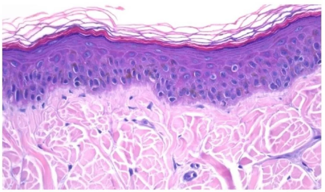
The EPIDERMIS is the superficial layer of the skin. It is not vascularized, i.e. it contains no blood vessels; it is not irrigated by blood. It is the first layer of the skin, but it is also made up of several distinct layers of cells. It defends the dermis against external aggressions. This is the thinnest layer constituting the skin.
The DERMIS lies under the epidermis and constitutes the second layer of the skin. It is not composed of several layers but mainly fibres and an amorphous substance containing different cells. It is the dermis that nourishes the epidermis and allows it to regenerate and maintain itself in good health. It provides the epidermis with everything it needs.
The SUBCUTIS lies deeper and supports the first two layers, also called panniculus or HYPODERMIS. Although it is NOT a layer of the skin, it is important to study it and to know how it works to fully understand how skin works.
Each of these three elements has fundamental interrelationships but to facilitate understanding, it is better to study them separately.
IMPORTANT
Although it is often said that the skin comprises of three layers, it actually comprises of two; the dermis and the epidermis, the third “layer” often called the hypodermis is not scientifically considered as a “layer” of the skin strictly speaking but it is necessary to know it to fully understand how the skin works.
The basement membrane
In addition to these three dermal elements, we must also mention the presence of the basement membrane between the epidermis and the dermis called "dermal-epidermal junction", "epidermal basal lamina" or "epidermal basement membrane". It separates the dermis and the epidermis and allows adhesion between its two dermal layers. It also serves as a physico-chemical barrier, a nutrient-oriented circulation and a component of the immune system. Deterioration of the basement membrane due to genetic problems, diseases, injuries, can lead to serious skin problems, some of which can be life threatening (skin cancer for example). The study of this membrane is only at the beginning, its deterioration is already associated with certain renal and dermal diseases.
In hairless areas, such as the paw pads and nasal planum, this junction is irregular because of epidermal projections that interdigitate with dermal papillae (e.g., rete ridges/also known as rete pegs), thus strengthening the epidermal-dermal attachment by providing resistance to shearing. In densely haired areas, the junction is smoother and has an undulating appearance as the epidermal-dermal attachment is strengthened by the hair follicles. (1)
The basement membrane zone also serves as a scaffold for migration of epidermal cells in wound healing and as an initial barrier to invasion of the dermis by neoplastic keratinocytes.
How can a groomer deteriorate the basement membrane?
In the medium or long term, it is entirely possible for the groomer to damage the basement membrane mainly by applying products that are too concentrated or in too large quantity. Therefore, it is imperative for any DAATA groomer to read and understand the labels of the cosmetic products used on the skin of the animals for which he is responsible.
Be sure to check the labels of your products and respect the recommended dilution rates and frequencies of use.
THE SKIN IS PERMEABLE TO EXTERNAL AGENTS IN MANY CASES
Normal intact skin has many natural defenses and barriers that render it impenetrable to most organisms and protect the body from a variety of other types of aggressions that include pressure, friction, mild mechanical trauma, external temperatures, UV light exposure, and chemical absorption.
The route by which an external agent gains entry into the body is called the portal of entry.
Many pathogens and chemicals can cause disease when entering the body via their specific portal of entry. A few pathogens, and chemicals, can penetrate intact normally functioning skin. Most of the time, the skin will only become an efficient portal of entry for microorganisms when the barrier is damaged by trauma, excessive moisture, heat or cold, or by disruption of the normal flora of the integument (during grooming for example). Intact skin with its waterproof barrier provides some protection against weak acids and alkali substances and water-soluble compounds, but as grooming remove this barrier, it exposes the skin to microorganisms and chemicals as well as to UV radiation (UVR) that can damage the skin by direct exposure especially if the body’s natural defenses, such as the hair coat and melanin pigments, are not present or are inadequate (1).
REFERENCES ON THIS PAGE
(1) The integument by Ann M. Hargis and Pamela Eve Ginn
(2) Schematic diagram of basement membrane structure of the skin. Keratinocytes attach to each other via desmosomes and to the basement membrane via hemidesmosomes. The projection of the basement membrane zone illustrates the multiple, interconnecting layers of this zone. The most superficial layer consists of the basal layer hemidesmosomes (keratin intermediate filaments and attachment plaques). The next layer, the lamina lucida, is an electron-lucent zone composed of the plasma membrane, subdesmosomal dense plate, and anchoring filaments. The deepest layer is the lamina densa, an electron-dense zone that consists of type IV collagen. Anchoring fibrils (type VII collagen), serve to attach the lamina densa and epidermis to the papillary dermis. The interconnecting layers of the basement membrane zone provide an important function in epidermal-dermal adherence, are the site of immune reactant deposition in cutaneous disease (see Table 17-11), and serve as a barrier to invasion by malignant epidermal tumors. ag, Apocrine glands; d, dermis; e, epidermis; hf, hair follicles; s, subcutis; sg, sebaceous glands. (Adapted from Kumar V, Abbas AK, Fausto N, et al: Robbins & Cotran pathologic basis of disease, ed 8, Philadelphia, 2009, Saunders; Rubin E, Farber JL: Pathology, ed 3, Philadelphia, 1999, Lippincott-Raven; and Elder DE: Lever’s histopathology of the skin, ed 10, Philadelphia, 2009, Lippincott Williams & Wilkins.)


