DAATA
02 The Epidermis
Functions + Definition | Keratinocytes | Melanocytes | Practice Test
The epidermis is the superficial layer of the skin, i.e. that which is exposed to the external elements. Unlike the dermis, it does not contain blood vessels, so it is not vascularized. It is the line of protection that protects the dermis against external aggressions. This layer is very dense and itself made of several layers of cells. This dermal layer is very thin. In hairy areas it is only 25 to 95 microns thick (less than one-tenth of a millimeter). The epidermis, however, is thick in the non-hairy areas of nose and pads. It consists of several types of cells. The epidermis (and the skin in general) is even thinner in the cat, being even thinner in the female than in the male in general from 12 to 45 micrometers (25 being the average in hairy areas).
Normal skin, haired, thorax, dog. The epidermis (arrow) in haired skin has an undulating surface but lacks rete ridges. The epidermis in haired skin has fewer nucleated cell layers than the epidermis in nonhaired (hairless) skin such as that on the nose and paw pads (see Fig. 17-2); thus, it is referred to as “thin” skin. Hair follicles (H), apocrine glands (A), and sebaceous glands (S) are present. The haired skin is thickest over the dorsal aspect of the body and on the lateral aspect of the limbs, and it is thinnest on the ventral aspect of the body and the medial aspect of the thighs. H&E stain. (Courtesy Dr. Ann M. Hargis, DermatoDiagnostics.)
Normal skin, hairless, paw pad, dog. The epidermis € in hairless (nonhaired) skin has more numerous nucleated cell layers and more abundant stratum corneum than the epidermis in haired skin; thus, it is referred to as “thick” skin. Note the dense zone of compact stratum corneum (SC) over the surface. The epidermis and dermal papillae in the superficial dermis (D) interdigitate to form rete ridges (arrows). The rete ridges strengthen the attachment between the epidermis and dermis. In the dog, the contour of the epidermal surface follows the epidermal ridges and thus is papillated. H&E stain. (Courtesy Dr. Ann M. Hargis, DermatoDiagnostics.)
The thickness of the skin differs according to the breeds, for example, the skin of the Labrador is thicker than that of the Pug, the Poodle or the Shitzu. (3)
In the table below, you can also see, for example, that the Shar-Pei is among the breeds with the thickest skin among those evaluated.
Figure 1 Measurements of skin thickness (epidermis plus dermis) determined from histologic examination of tissue sections and evaluation of ultrasonographic images obtained from 26 clinically normal dogs.
It also differs according to the anatomical area. On the diagram presented below, we can see a much greater thickness at the level of the interdigital spaces. The skin in the throat is among the thinnest, as well as under the armpits (Axilla). It can also be noted that the stratum corneum is particularly thin in these areas as well as in the ears.
Figure 2: Source: Theerawatanasirikul, Sirin & Suriyaphol, Gunnaporn & Thanawongnuwech, Roongroje & Sailasuta, Achariya. (2012). Histologic morphology and involucrin, filaggrin, and keratin expression in normal canine skin from dogs of different breeds and coat types. Journal of veterinary science. 13. 163-70. 10.4142/jvs.2012.13.2.163.
Keratinocytes
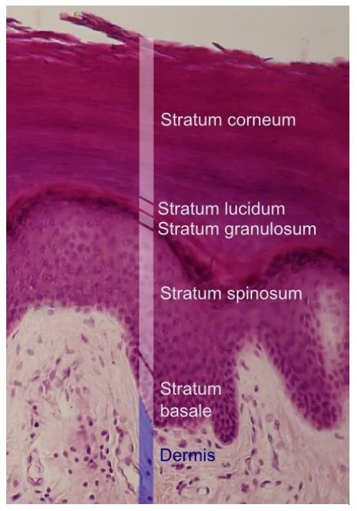
The epidermis is divided into layers based on the morphology of the keratinocyte, the major cell type of the epidermis. The epidermis of haired skin consists of four basic layers: stratum corneum (sc), stratum granulosum(sg), stratum spinosum(ss), and stratum Basale (sb). The epidermis of hairless skin consists of five layers; the fifth layer is the stratum lucidum (sl), which is located between the stratum granulosum and stratum corneum. Keratinocytes originate from germinal cells in the stratum Basale of the epidermis, ascend through the layers of the epidermis, changing in appearance and other characteristics in each layer until they reach the stratum corneum as fully keratinized, dead corneocytes. Keratinocytes are continuously shed from the stratum corneum. The transit time for a keratinocyte from the stratum Basale to shed in the stratum corneum is approximately 21 to 28 days, although this time can be accelerated in some disorders such as primary seborrhea characterized clinically by scaling. So, as you can see, the skin is living organ renewing constantly.
Schematic diagram of the structure of the skin. A, the skin is composed of epidermis €, dermis (d), and subcutis (s), with adnexa consisting of hair follicles (hf), sebaceous glands (sg), and apocrine glands (ag). B, This projection of the epidermis demonstrates the progressive upward maturation of basal cells (bc) in the stratum Basale (sb) through the stratum spinosum (ss), stratum granulosum (sg) into the cornified squamous epithelial cells of the stratum corneum (sc). Melanocytes (m), midepidermal dendritic Langerhans’ cells (lc), and Merkel cells (mc) are also present. The subjacent dermis contains small vessels (v), fibroblasts (f), perivascular mast cells (pmc), and dendrocytes (dc), potentially important in dermal immunity and repair. (Adapted from Kumar V, Abbas AK, Fausto N, et al: Robbins & Cotran pathologic basis of disease, ed 8, Philadelphia, 2009, Saunders; Gawkrodger DJ: Dermatology: an illustrated colour text, ed 2, New York, 1997, Churchill Livingstone.)
The outermost layer of the epidermis is the stratum corneum, which consists of many sheets of flattened, keratinized cells termed corneocytes. Keratin is an intracellular fibrous protein that is in part responsible for the toughness of the epidermis, enabling the epidermis to form a protective barrier.
Underneath the stratum corneum is the stratum granulosum, which consists of effete cells. The name granulosum comes from the granule’s shapes of the cells that compose this layer.
In non-haired skin, the stratum corneum and stratum granulosum are separated by an additional layer of compacted, fully keratinized cells, the stratum lucidum, best seen in the paw pad.
Deep to the stratum granulosum is the stratum spinosum, a layer of polyhedral-shaped cells attached to one another by desmosomes. During fixation and processing for microscopic examination, the cells of the stratum spinosum contract, except for the desmosomal attachments. These attachment sites create the appearance of “spines” or intercellular bridges, leading to the name of this layer.
The innermost layer of the epidermis is the germinal layer or stratum Basale sometimes referred as stratum germinativum, which consists of a single layer of cuboidal cells resting on a basement membrane. Intermixed within the basal cell layer are melanocytes, Langerhans’ cells, and Merkel cells.
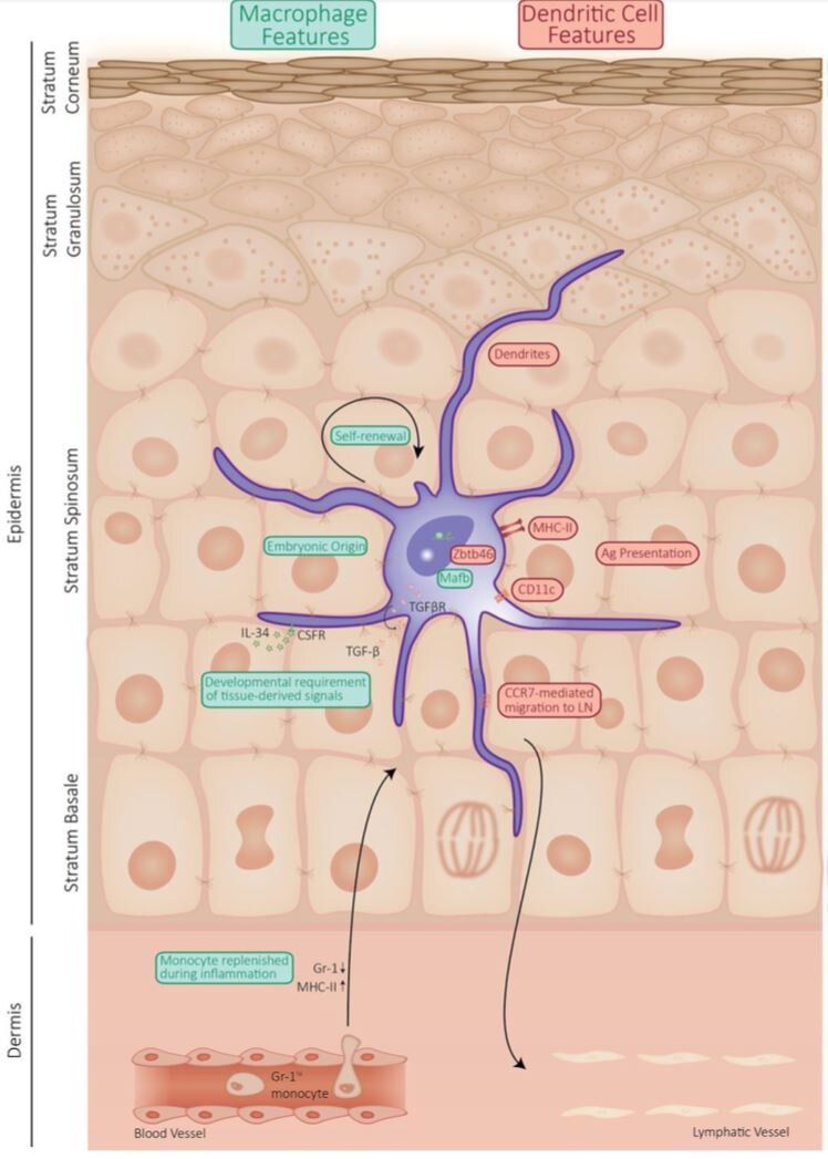
Langerhans cells recognize pathogens and influence the immune response. There are situated between the keratinocytes of the basal layer and the thorny layer, but mainly in the thorny layer. They make up 3–5% of all nucleated cells in the epidermis and occupy the stratum spinosum. Langerhans cells are, due to their location within stratified epithelia, part of the first line of defense to pathogens present in the environment.
(This detailed diagram is there just to help you identify the location and size of Langerhans cells in the epidermis. The technical and scientific terms in this diagram can be ignored and will not be the subject of questions during your final exam.)
Source: Langerhans Cells: Sensing the environment in Health and Disease Julie Deckers, Hamida Hammad and Esther Hoste
Merkel cells are nerve receptors that play an important role in tactile sensation (4). They are the specialized skin epithelium light touch sensors. There are all placed in the basal membrane and below and linked to a nerve ending (5). They play an important role in nerve functioning and act as receptors. Merkel’s cells may have other functions, such as influencing cutaneous blood flow and sweat production (via the release of vasoactive intestinal peptide), coordinating keratinocyte proliferation, and maintaining and stimulating the stem cell population of the hair follicle (hence controlling the hair cycle) (6).
Melanocytes allow pigmentation of skin and hair. They synthesize pigments called melanin that colour the skin.
Melanocytes
UNDERSTANDING SKIN PIGMENTATION
There are two types of melanocytes that pigment skin and hair, that is, there are only two colours of pigment. All the colours of dogs and cats come from these two colours. (7,8)
EUMELANIN – BLACK PIGMENT
Eumelanin is basically a black pigment. All black areas of a dog are the result of the production of Eumelanin by melanocytes. However, some genes alter the colour of the black pigment which can be brown, blue or very pale brown. In dogs, the presence of a modifying gene will turn the entire black colour into brown, gray or pale brown. This is a dilution of black for the blue and pale brown and a change in the structure of the pigment for the brown.
It is quite possible, even if it is rare, to have a dog (and more often a cat) with solid pigments and diluted pigments at the same time. The most famous in dogs is called Merle.
The eumelanin also colours the eyes and the nose. Depending on the type of eumelanin that the dog can produce, the nose will be black, brown, blue or pale brown. Mostly, dogs and cats have brown eyes. Here, the iris is coloured by superposition of layers of black pigments. When the body is not genetically capable of producing enough eumelanin, the colour of the eye is clearer. The eye can then be light brown, yellow gold or blue.
PHAEOMELANIN
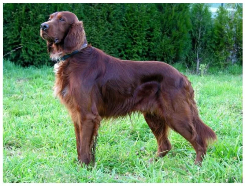
Phaeomelanin is a red pigment. The term “red” covers absolutely all the deep red variations of the Irish Setter with the clearest cream, including, the sand, the fawn, the orange. This melanin is only produced in the hair. It is not produced in the eyes or on the nose. So, the gene that affects the intensity of the red colour, will not affect the colour of the eyes or nose.
WHITE
White is not a colour; it is an absence of colour. Therefore, animals with white hairs do not produce eumelanin or phaeomelanin.
To be more precise, the genes do contain a colour, but it does not stand out because a genetic code tells the body not to colour the hair. Thus, white dogs and white cats have a genetic colour, but we cannot know which one. From the point of view of grooming this raises the problem of whether it is a coat with diluted colour because the grooming and maintenance is then more complicated.
THE OTHER WHITE
But there is also a second type of white, caused by the dilution of red pigments. Sometimes the dilution is so important that the hair looks white. In these animals, you will see as a cream shadow in the coat indicating the very slight production of pigments.
This type of white, generally does not concern eumelanin, therefore the black, brown, blue or pale brown areas on the hair will keep their colour, as well as the eyes and the nose.
ALTERNATING PIGMENTATION
Pigments determine the colour of hair and skin and they distribute a certain amount of eumelanin and phaeomelanin. They order certain cells to produce one and others to produce the other.
Sometimes, genes direct cells to produce no melanin at all. Sometimes the genes can order to change the type of melanin produced, that is from eumelanin to phaeomelanin.
This means that the hair will produce coloured bands along the strand.
For example, it produces black pigments for a while, then red pigments, then it comes back to black and so on. This is the “agouti”. It is a very common colour in rabbits or wild mice, as well as in many other wild mammals because it brings a natural camouflage.
This little rodent is an agouti and gave his name to the agouti coat in cats and dogs.
Notes on the genetics of agouti in cats
Cats with A/A genotype will have agouti banded hair. They will transmit this agouti variant to all of their offspring, and all of their offspring will have banded hair.
Cats with A/a genotype will have agouti banded hair. They will transmit this agouti variant to 50% of their offspring, and the offspring of an A/a genotype cat can be agouti or non-agouti depending on the genotype of the mate.
Cats with a/a genotype will have self-colored (solid) hair. If bred to another non-agouti cat (a/a genotype), all offspring will also have non-banded, solid-coloured hair.
In this photo, a white poodle has been clipped very short to allow the skin to tan and create a deep contrast with the white coat.
IMPORTANT TO KNOW
The more pigmented the skin, the more resistant it is to ultraviolet.
It should be noted, from this perspective, that the cat produces very few pigments in the skin. Almost all the coloring is sent in the hair. Clipping a cat will make it even more vulnerable to ultra-violet than a dog. Similarly, clipping is even more dangerous for dogs whose skin is less pigmented and the production of melanocytes is not enough to “tan” the skin. That is to say, all the light coats and/or skins.
WHY IS PIGMENTATION IMPORTANT FOR THE GROOMER?
Knowing the different colours and how they work is particularly important for the groomer specially to determine the right grooming schedule of a pet. Indeed, according to each colour, the texture and thickness of the hair will be different. The solid black and red colours produce thicker hairs with a smooth surface. The solid brown colour produces a slightly finer and slightly wavy coat. While the diluted colours produce an even finer and much wavier coat. Due to this wavy structure and the lightness of the hairs, they knot faster. Thus, brown coats knot faster than black coats and diluted coats knot faster than solid coats. So, with the same maintenance and lifestyle, a black dog needs less frequent grooming than a blue dog.
INDICATIVE GROOMING SCHEDULE ACCORDING TO COAT COLOURS
o Diluted coats should be groomed every 4 weeks approximately.
o Brown coats should be groomed every 6 to 8 weeks approximately.
o Black coats should be groomed every 8 to 10 weeks.
These are only indicative and will vary according to the type of coat, the pet’s lifestyle, and the length of the hair.
Being able to determine the proper grooming schedule of each pet and explaining the reasons to the customer is an essential element of professionalism for the groomer. The DAATA method will allow you to scientifically justify your decisions to your customers and will allow you to exercise your profession in better conditions. This will result in better quality grooming and an improvement in the condition and welfare of the pets you care for.
WATCH NATHALIE’S LECTURE
REFERENCES ON THIS PAGE
(3) Source: Theerawatanasirikul, Sirin & Suriyaphol, Gunnaporn & Thanawongnuwech, Roongroje & Sailasuta, Achariya. (2012). Histologic morphology and involucrin, filaggrin, and keratin expression in normal canine skin from dogs of different breeds and coat types. Journal of veterinary science. 13. 163-70. 10.4142/jvs.2012.13.2.163.
(4) Anatomical Mapping and Density of Merkel Cells in Skin and Mucosae of the Dog GUSTAVO A. RAMIREZ, FRANCISCO RODRIGUEZ,OSCAR QUESADA,PEDRO HERRAEZ, ANTONIO FERNANDEZ,ANDANTONIO ESPINOSA-DE-LOS-MONTEROS Unit of Histology and Veterinary Pathology, Institute for Animal Health, Veterinary Col-lege, University of Las Palmas De Gran Canaria, Campus Universitario Cardones, Arucas, Las Palmas 45413, Spain
(5) Laura B. Stokking PhD, DVM, in Small Animal Dermatology Secrets, 2004
(6) Danny W. Scott DVM, William H. MillerJr. VMD, in Equine Dermatology, 2003
(7) Pigment Intensity in Dogs is Associated with a Copy Number Variant Upstream of KITLG Kalie Weich, Verena Affolter, Daniel York, Robert Rebhun, Robert Grahn , Angelica Kallenberg and Danika Bannasch
(8) See also : Sheila M. Schmutz, Ph.D., Professor, http://homepage.usask.ca/~schmutz/dogcolors.html


