DAATA
03 Hair and Coat
Introduction | The Follicle | Anatomy of the Hair | Pilar Cycle | Shedding | Skin Glands | Practice Test
The epidermis synthesizes the hair through the hair follicles which are extensions of the epidermis. These are small bags. Hair follicles are created in the embryonic stage. If a follicle is destroyed it cannot be regenerated by the body. It is therefore essential to maintain the skin and the hair to preserve the follicular capital.
In dogs and cats, a follicle is compound, meaning that many hairs come out of the same follicle.
Hair follicles are usually positioned at a 30-60-degree angle to the skin surface. Although hair length, thickness, density and colour vary between individuals and especially between breeds, all hairs have the same basic structure (Paus et al., 1999).
THE FOLLICLE
The hair follicle consists of three distinct parts:
From the top (skin surface) to the bottom:
• The infundibulum,
• The isthmus
• The deep part also called hair bulb.
The infundibulum consists of the segment from the entrance of the sebaceous duct to the skin surface. The isthmus consists of the segment between the opening of the sebaceous duct and the attachment of the arrector pili muscle. The inferior segment extends from the attachment of the arrector pili muscle to the dermal hair papilla (Miller et al., 2013a).
The follicle is also comprised of different parts:
The Ostium is the hair exit orifice. All hairs in a follicle come out of the same ostium. The hair and inner root sheath are formed by pluripotent stem cells in the hair matrix. The outer root sheath represents a downward extension of the epidermis (Miller et al., 2013a). The sebaceous gland is one of the main cutaneous glands, producing sebum. Thedermal papilla lies in the deep part of the follicle.
Figure 3 Schematic depicting the five major components from the hair follicle and the three anatomic segments.
THE DERMAL PAPILLA
It is very important to understand what the dermal papilla is and how it works. The dermal papilla is attached to a blood vessel and nerves. This vessel brings the necessary nutrients that will create the cells forming the hair. During the growing phase, the hair is attached to the papilla. Removing this hair (for example by pulling the wrong hair during hand stripping or brushing too hard or using a comb to remove the matts) rips off the papilla and creates a micro wound and therefore, an open door to the blood system. This can lead to an infection of the follicle and creates further skin issue. The trauma caused to the follicle can lead to inflammation and lead for example to inflammation of the sebaceous gland which will increase the production of sebum. Too much sebum will lead to more oily skin and hair, an imbalance of the skin flora, engorgement of the follicle which can lead to the stopping of hair regrowth, blackheads, pustules, folliculitis, etc. The pet may also emit bad odours.
DETAILS OF A FOLLICLE
On the picture below you can see the top view of the infundibulum (b). The biggest white hole is the “bag” of the primary hair. The medium size white hole are the bags of the primary lateral hair and the smallest ones are those of the secondary hair.
NOTE
It is the skin that determines how healthy a hair is. An unhealthy skin creates unhealthy hair. You must preserve the skin if you want to be able to work with a beautiful hair and therefore obtain the best grooming results.
ANATOMY OF THE HAIR
It consists of a root and a stem. The root is in the depth of the skin, it is not visible. The stem has a free end that protrudes from the skin surface. The hairs consist of different proteins and especially Cystine.
The hair consists of three parts from the inside to the outside:
• The Medulla is in the center of the hair. These are flattened cells from top to bottom. They are solid near the base of the hair, but the higher in the hair, the more it contains air and glycogen vacuoles.
• The Cortex consists of cornified cells elongated parallel to the major axis of the hair. It contains the pigments that give colour to the hair and represents 1/6th to ¼TH of the thickness of the hair.
• The Cuticle is composed of flattened, cornified, anucleate cells, organized like tiles on a roof. They are all flat and very close to each other. They give the hair its soft and silky appearance to the touch.
THE FUR HAS MANY ROLES
- Protection (against the rain, the sun)
- Social recognition (colour and smell),
- Thermal regulation (undercoat helps regulating heat and cold)
- Sense organ (specialized hair).
- Defense against predators (camouflage) as seen previously with the Agouti colour for example.
THE PROTECTION ROLE OF THE FUR IS PLAYED BY THE COMPOUND FOLLICLE
Thanks to the compound follicles, the coat of dogs and cats protects the animal against sun, rain, hot and cold.
Dogs and cats have a compound coat, meaning that several hairs come out of the same hair follicle. To be more precise, the follicle comprises of several bags of different sizes according to the type of hair growing inside. Each bag carries a hair and all the hair coming out of all these bags come out of the same “ostium”.
Generally, in dogs, each hair follicle contains a primary (largest) hair associated with 2 to 5 smaller primary lateral hair and up to 20 secondary hair.
The primary hair and primary lateral are larger:
• The primary hair or jar hair is rigid, it covers the entire skin surface and are a protection against rain.
• The primary lateral hair (or guard hair) is moderately supple with a different orientation than that of the undercoat. It allows better thermal insulation.
Each primary hair arises from its individual pore and has sebaceous glands, sweat glands and an arrector pili muscle.
The secondary hair, smaller, make up the down or undercoat and help maintain body temperature. It is fine and flexible. Secondary hairs are only accompanied by sebaceous glands and emerge through a common pore.
The coat thus formed ensures thermoregulation of the skin and therefore of the body.
- The primary hairs play the role of the roof over the other hairs, protecting them from the rain and preventing water from reaching the skin which would cause a drop in skin temperature.
- The lateral primary hairs, thanks to their texture and their orientation, keep the secondary hair in order, it will thus limit the formation of knots and allow air to enter the coat in order to lower the skin temperature. The more secondary hairs there are, the less air will enter and the less secondary hairs the more air will enter.
- Primary hairs also contain most pigments which, among other things, protect the skin against ultraviolet rays.
- The secondary hairs have the role of heating blanket. Depending on their quantity, the skin is kept warm.
A good balance between all types of hair allows optimal thermoregulation.
DIFFERENT TYPES OF COAT
Not all coats contain the same proportions of primary and secondary hair. The differences in coats are first genetic and, depending on the breed, the distribution of each type of coat will be different. Depending on each individual then and internal or external factors, the distribution will also change from one individual to another.
Coat differences by breed (or individual) depend on the predominant type of hair.
In the German Shepherd for example, which has a "classic coat", primary and secondary hairs are developed evenly. And depending on the season, the distribution between primary hairs and secondary hairs will change so that thermoregulation is carried out properly.
In short and coarse hair breeds, primary hairs are developed and there are few secondary hairs. Its breeds are generally terriers which need more protection against water and against external physical aggressions (entering brambles, burrows, etc.).
In short and thin hair breeds, there are many secondary hairs and the primary hairs are small.
In long-haired breeds, most often they have a significant proportion of secondary hairs. The Coton de Tulear, the Bichon Frisé, etc, are a few examples. The poodle coat is mainly made of secondary hair.
Some others long haired breeds have a well-developed primary coat with a moderate amount of undercoat (the Shih Tzu for example).
In the same breed, the type of coat can vary greatly. For example, in the Shih Tzu standard, it is stated that this breed should only have a little undercoat. Some individuals do have a huge amount of undercoat. On the contrary, the powder puff Chinese crested is supposed to have a huge amount of undercoat, but certain individuals do not develop a lot of it. You might even see some differences on certain parts of the body, some developing a lot of undercoat and some other parts developing only a little.
GENETIC TRANSMISSION
Many features of the coat are transmitted by an autosomal recessive gene. An autosomal gene is a gene other than sexual (neither X nor Y, therefore). Recessive means that both parents must transmit the gene for it to come out.
For example, the "brown" colour from chocolate to bronze in the Newfoundland dog, the Tibetan terrier and the Shar-peï, the longhair character in the Alaskan Malamute, the Corgi, the Hungarian Braque, the German Shepherd, the Mastiff, the Belgian Shepherd, the Weimaraner, the dilutive character of the Great Dane, the Shar Pei, the Azawakh, the Staffordshire Terrier, the Bull Terrier, the Greyhound, the Belgian Shepherd, the Whippet.
The pilar cycle also is determined by genetic transmission.
THE PILAR CYCLE
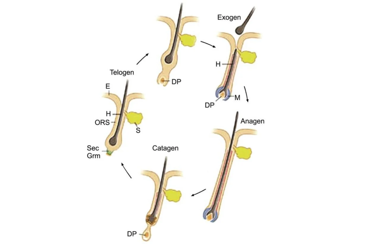
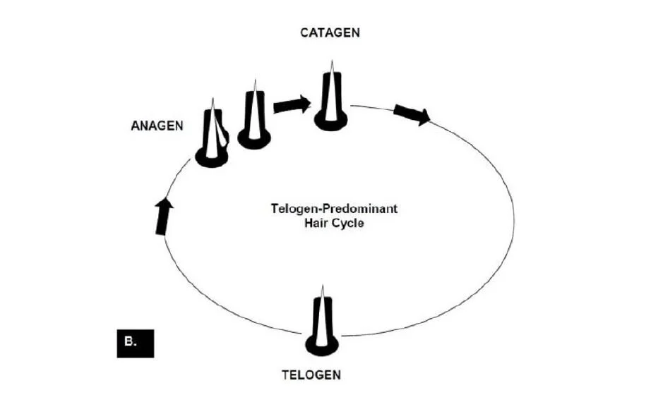
The hair cycle is divided into three main distinct phases, themselves interspersed with intermediate phases. The first one is the anagen phase is the growth phase of the hair. The intermediate phase or catagen is the phase of degeneration. Finally, the telogen phase is also called the rest phase. In this last phase, the hair does not evolve anymore, it waits for another hair to enter the anagen phase and expel it from the follicle. Once a new phase starts, the hair is expelled of the follicle, this is called the exogen phase. This sequence of anagen, catagen, telogen and exogen is defined as the hair growth cycle.
Figure 4 Schematic depicting the different hair follicle stages of the hair follicle cycle. E: epidermis, ORS: outer root
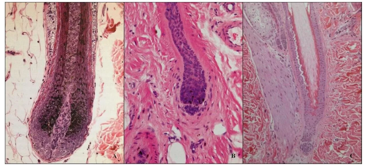
The histologic appearance of the hair follicle varies with the stage of the hair follicle cycle (Miller et al., 2013a; Al-Bagdadi et al., 1979; Mecklenburg, 2009a). The anagen hair follicle extends into the deep dermis and often the subcutis. It is characterized by a well-developed spindle shaped hair dermal papilla capped by the hair matrix. Matrix cells can show mitotic activity and are often heavily melanized.
The catagen hair follicle is characterized by retraction towards the surface. The hair follicle epithelium shrinks from the hair bulb to just below the entry of the sebaceous gland duct. The dermal papilla is round to oval shaped. The proximal hair shaft is pigmented, melanogenesis ceases and mitotic activity stops. The inner root sheath is partially replaced by trichilemmal keratinization.
The telogen hair follicle is reduced to about one third of its former length. The dermal papilla is small and is separated from the matrix cells. There is no melanin and no mitotic activity. The inner root sheath is absent. A distinct club hair has now formed that demarcates the isthmus region of the follicle (Miller et al., 2013a; Al-Bagdadi et al., 1979; Mecklenburg, 2009a). Directly below the isthmus there is a secondary hair germ. These cells will produce a new hair follicle once a new anagen phase is initiated (Mecklenburg, 2009a). The small and condensed dermal papilla is found directly below this secondary hair germ. In exogen hair follicles the hair is shed from its follicle (Miller et al., 2013a; Higgins et al., 2009). This stage of the hair follicle cycle has received little attention to date.
Figure 5 Histologic pictures of an anagen (a), catagen (b) and telogen(c) follicle. A. Anagen follicle. Note dermal papilla, matrix with melanocytes. B. Catagen hair bulb with dermal papilla, prominent basement membrane and epithelial strand. C. Note telogen hair follicle with club hair and trichilemmal keratinization (white arrow). Prominent basement membrane separating dermal papilla (black arrow) and secondary germ. Adapted from Mecklenburg, 2009.
Figure 6 Schematic diagram of the hair cycle. A, Anagen. During this growing stage, hair is produced by mitosis in epithelial cells covering the apex of the follicular papilla (dermal papilla), which is enveloped by hair matrix cells at the hair bulb. B, Early catagen. In this transitional stage, a constriction occurs at the hair bulb, and the hair shaft above this becomes a “club hair.” C, Catagen. The distal follicle becomes thick and corrugated and pushes the hair outward. D, Telogen. This is the resting stage in which the follicular papilla separates and an epithelial strand shortens to form a secondary germ. E, Early anagen.
The secondary germ grows down to enclose the follicular papilla, and a new hair bulb forms. F, Exogen. This refers to the shedding of the old hair shaft. G, Anagen. The hair elongates as growth continues. Note that the anagen hair bulbs are located deeper in the dermis than the catagen and telogen hair bulbs. (Adapted from Scott DW, Miller WH Jr, Griffin CE: Muller and Kirk’s small animal dermatology, ed 6, Philadelphia, 2001, Saunders.)
SEVERAL FACTORS DETERMINE THE LENGTH OF THE CYCLES
Genetic Factors
From a cycle length perspective, there are two distinct breed groups in dogs and only one in cats.
The first group are the anagen breeds as they have an extremely long growth phase such as Yorkshire Terriers. They do not have a pre-determined length of hair. The hair will grow continuously and can be extremely long if properly maintained.
Most of the breeds are telogen breeds, they have a genetically predetermined length of hair. Once this length is reached, the hair stops growing and enter into telogen phase. The telogen phase is then very long and will last until a new anagen phase starts. This new anagen phase usually starts with a new shedding. In some breeds, such as the Nordic breeds (Husky, Malamute, etc.), the telogen phase can be very long and lasts up to 12 years approximately (the whole life of the dog) (9).
OTHER FACTORS
Cycles also follow external factors, such as seasonal change, photoperiod, diet. Finally, the duration of the phases also depends on internal factors such as hormones, vascularization or growth factors. Stress can also be a factor.
Photoperiod acts via the hypothalamus, pituitary, and pineal glands, which secrete tropic hormones, such as melatonin and the gonadal, thyroid, and adrenal hormones, that also influence hair growth. Some hormones, such as thyroid and growth hormone, stimulate hair growth, whereas excessive levels of estrogen or glucocorticoids suppress hair growth. The cells in the follicular papilla (sometimes called dermal papilla) regulate epithelial growth and are probably the target of the tropic hormones. Nutrition and health status have a significant influence on hair growth and quality. Hair is largely composed of protein. Thus, diets low in protein or disease states associated with severe protein loss result in poor quality hair coat. Animals in poor health have larger numbers of hair follicles in telogen, and because telogen hairs are more easily shed than anagen hairs, animals in poor health tend to shed more heavily than healthy animals. Also, in disease states, cuticle formation can be defective, resulting in a dull or dry hair coat. It is also thought that the hair cycle is influenced by the nervous system. Evidence of such is implied by the abundant nerve supply of the hair follicle, the high density of Merkel cells in the hair follicle epithelium, and facts that keratinocytes express several neurotransmitter and neuropeptide receptors whose stimulation can alter keratinocyte proliferation and differentiation. Indirect influence by autonomic nerves could also be mediated by alteration of hair follicle blood flow and thus of oxygen and nutrient supply.
The catagen phase generally lasts about ten days.
IMPORTANT TO UNDERSTAND
Each follicle has an independent cycle of the others, so each one is at a different stage in the growth cycle. Therefore, a dog or cat is never completely naked at times.
SHEDDING
When certain factors cause a new anagen phase on a greater or lesser proportion of the coat, the animal sheds.
There are several types of shedding:
The “mosaic shedding”, a gradual renewal of the coat and permanently in different areas. Therefore, the animal constantly loses its hair throughout the year. This type of shedding is found mostly in animals that live indoors and do not have to undergo photoperiod changes.
The second type of shedding is called “main shedding”.
First, spring shedding with the formation of a less dense coat to overcome the arrival of higher temperature and an increase in exposure to light.
Second, the autumn shedding with the constitution of a denser coat, a winter coat to palliate the decrease of the sunshine and the temperatures. During these two main phases, there is maximum hair growth in late spring and minimal growth during the winter. Animals that live outdoors shed twice a year while animals that live indoors shed throughout the year.
In summer, with variations between breeds, an average of 30% of primary follicles and 50% of secondary follicles are at rest (dead hairs in telogen phase no longer grow). In winter, 75% of primary follicles and 90% of secondary follicles are at rest.
In some breeds there is also a partial or complete shedding during the period of lactation or gestation, this shedding is called effluvium.

CAT'S COAT SPECIFICITIES
Short-haired cats have primary hairs measuring approximately 4.5 cm for 15 cm in long hairs. Short-haired phenotypes are said to have a wild-type phenotype (L) and are more numerous than long-haired phenotypes (1). All cats are telogen breeds. The types of hair are similar to dogs. There are exceptions such as Rex breeds where a genetic mutation causes curly / twisted hair to form. In Devon Rex, secondary hairs are like primary hairs. In Cornish Rex, there is no primary hair at all. In the cat, each follicular unit also contains 2 to 5 primary follicles for 5 to 20 secondary follicles. The difference with the dog’s coat lies in the density of the coat which is much higher than that of the dog with 600 to 1800 hairs per cm²! In the dog it is around 1000 hairs for 6 cm². Unlike the dog, the colour of the cat depends almost solely on his hair. Meaning that there is only a little protection in the skin. Clipping a cat short, leaves him with no protection whatsoever against the sunrays. It is important to educate your customers and colleagues about the fact than a cat should only be clipped as a last resort. If clipped, the owner must take the necessary precautions to protect his pet against the sun and minimize his exposure to direct sunlight until the hair has regained enough length.
SKIN GLANDS
Most skin glands in dogs and cats are associated with hair follicles and hair (whereas in humans they are associated with the skin surface). We can mention the two main glands:
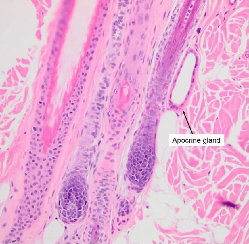
The sweat glands (which secrete sweat and odors), also called Apocrine glands, that reduce body temperature by evaporation. Sweating is a very undeveloped phenomenon in dogs, a little more in cats. They therefore have almost no effect on the thermoregulation provided by carnivores by respiration. On the other hand, these glands also synthesize various odoriferous substances that probably have a major role in sexual identification and probably also in social interactions.
FIGURE 7: DERMAL ADNEXA, DOG. APOCRINE GLANDS ARE ASSOCIATED WITH HAIR FOLLICLES, AND AS SUCH, ARE REFERRED TO AS EPITRICHIAL GLANDS.
The sweat glands (which secrete sweat and odors), also called Apocrine glands, that reduce body temperature by evaporation. Sweating is a very undeveloped phenomenon in dogs, a little more in cats. They therefore have almost no effect on the thermoregulation provided by carnivores by respiration. On the other hand, these glands also synthesize various odoriferous substances that probably have a major role in sexual identification and probably also in social interactions.
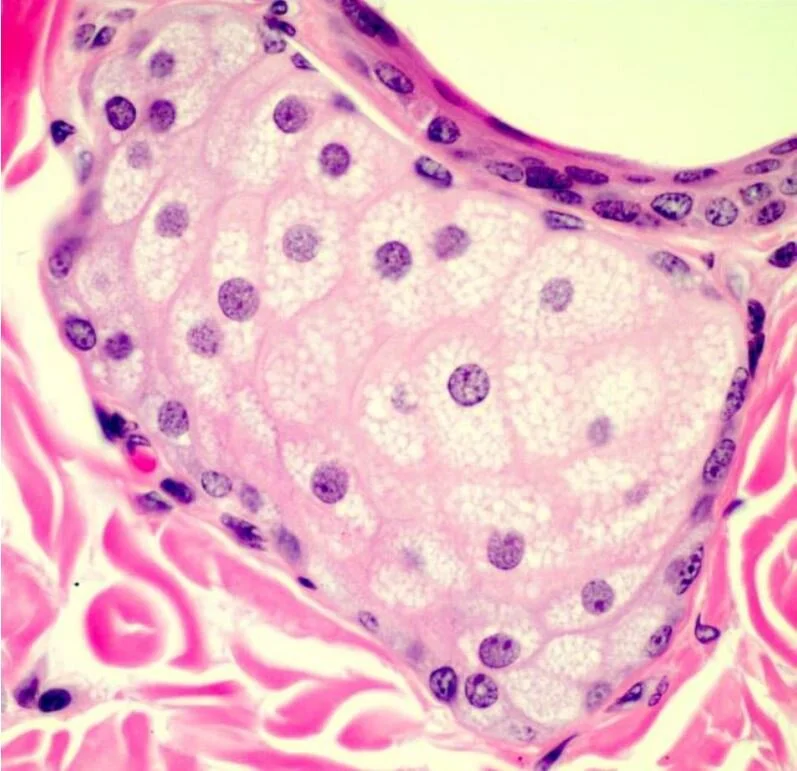
FIGURE 8 VETERINARY HISTOLOGY BY RYAN JENNINGS AND CHRISTOPHER PREMANANDAN
The sebaceous glands produce sebum, a secretion rich in fat that integrates with the superficial stratum corneum and provides protection and flexibility to the skin. Sebum also protects against the development of certain microbes. It sheaths the entire surface of the hair and gives it its lustrous and smooth appearance. It provides protection against external aggression. The glands are present on all the skin except the nose and the pads. A dysfunction of these glands causes the phenomenon of seborrhea and bad smells.
FIGURE 9.. THE SINUS HAIR (IEH) IS A VIBRISSAE; THE LANUGO HAIR (RIGHT) IS A NORMAL BODY HAIL'. NOTE THE SIZE OF THE SENSORY NERVE ENTERING THE VIBRISSALROOT TO THE LEFT, PLUS THE SECOND NERVE ENTERING THE TOP RIGHT. REPRINTED FROM ANDRES, K. H., AND VON DURING, M. "MORPHOLOGY OF CUTANEOUS RECEPTORS" IN HANDBOOK OF SENSORY PHYSIOLOGY, VOL. 2, P. 24: SPRINGER.VERIAG, NEW YORK, 1973.
The coat is also a sense organ thanks to specialized hairs
The fur consists of other hairs with a specialized role: the vibrissae present on the face and the tylotriches hair scattered on the whole body.
Anatomically, vibrissae are constructed much differently from other body hair. They are thicker, stiffer, rooted in erectile tissue and placed so as to act as levers on the nerves serving them. Vibrissae are much more heavily innervated than other body hair; that is, more nerve fibres serve each vibrissae. The vibrissae in dogs are served by the trigeminal nerve, the largest of the 12 pairs of canine cranial nerves larger than the optic nerve, auditory nerve, or olfactory nerve. (10)
They have an important role in the dog's life, especially for movement and recognition between individuals (social function). It is presumed that they have many other important roles in the life of the animal.
WATCH NATHALIE’S LECTURE
REFERENCES ON THIS PAGE
(9) (Tobin, 2007; Lloyd et al., 2012; Mecklenburg, 2006).
(10) "WHISKER" TRIMMING IN SHOW DOGS: a harmless cosmetic procedure or mutilation of a sensory system? by Thomas E.McGill, Ph.D. Hales Professor of Psychology WilliamsCollege Williamstown, MA


